Ultrasound
Ultrasound, most commonly associated with pregnancy, is not something you may expect to hear at a veterinary office. However, due to improvements in technology, an ultrasound may be something your veterinarian recommends to help diagnose your pet for a number of potential ailments.
An ultrasound is a non-invasive procedure similar to an x-ray. It works by sending sound waves through tissue and recording the waves as they are reflected back. Those reflections are then transformed into images of organs and other objects for your veterinarian to study. In simplest terms, an ultrasound produces a moving picture of an organ or body part as it is actually functioning.
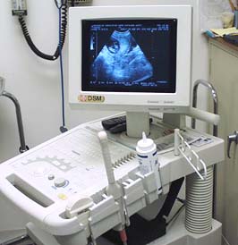
Veterinary Ultrasound Machine
Most commonly used as a diagnostic tool to evaluate diseases of the heart, liver, pancreas, kidney, intestine, spleen, urinary bladder and other organs located in the abdomen, your veterinarian is able to learn valuable information about the health of these organs. Since the pet is usually on his or her back for an ultrasound procedure, sedation or short acting anesthesia may be required. Regardless if sedation is used, ultrasound is an out-patient procedure, usually allowing your pet to go home the same day.
The benefits of ultrasound are enormous. Diseases that would otherwise go undetected can be diagnosed early. If a biopsy is needed, it can be accomplished during an ultrasound. An ultrasound can also replace an exploratory surgery, which can sometimes lead to more serious complications.
Although there are other parts of the body that can be studied with ultrasound, abdominal and cardiac ultrasound are the most common in veterinary medicine.
Abdominal Ultrasound
Abdominal ultrasound is used to evaluate pets with symptoms such as vomiting, diarrhea, straining to urinate or urinating blood. It can also be helpful in cases of reproductive abnormalities, weight loss and to detect early pregnancy. When physical examination and blood tests indicate a problem with a particular organ, an ultrasonic examination can provide additional information or even a diagnosis.
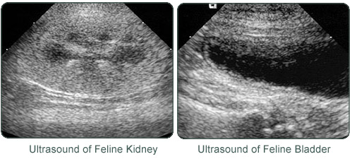
In order for the ultrasound to produce the best possible picture, a small amount of fur needs to be shaved from the abdomen. After the fur is shaved, the examiner places a probe on the skin of the abdomen and moves it across the surface to examine the organs or regions of interest. An ultrasound can identify organ abnormalities, abdominal masses, tumors, fluid and abnormal lymph nodes.
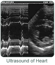
Echocardiography
An ultrasound of the heart is more commonly known as an echocardiogram. Defined as an ultrasonic examination of the heart, the procedure itself is very similar to that of an abdominal ultrasound.
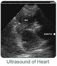
An ultrasound allows the veterinarian to see inside your pet's heart. The functioning of the heart valves, the thickness of the heart muscle and the contractions of the heart can all be assessed. Along with a diagnosis, an echocardiogram also allows the veterinarian to determine the best treatment plan for your pet's condition.
Ultrasound has become a very useful diagnostic tool in veterinary medicine. To a large extent, ultrasound has replaced exploratory abdominal surgery. Along with radiography, ultrasound can also be used to diagnose and treat most heart diseases that occur in dogs and cats.